
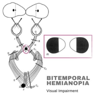
Bitemporal Hemianopia – Visual Impairment: Bit-temporal hemianopsia is partial blindness in the outer half of both the left and the correct fields of vision absent. It is often linked to the optical chiasm lesion. It is the intersection between the left and the right eyes of the optic nerves at the hypophysis.
The visual impairment is often manifested as bitemporal hemianopsia or superior quadrantanopia following supra meningiomas (Bejjani et al., 2002). This deficiency pattern is supposed to be caused by weak optic chiasm compression (Walsh, 1992). Peritorcular meningioma operation might lead to temporary or permanent loss of vision. These damages are tough to recover, and no specific technique is available. Two main options are optical devices and compensatory training.
Bitemporal Hemianopia – Visual Impairment
Causes of Bitemporal Hemianopia
The optic chiasm almost damages bitemporal hemianopia. It may result in several injuries, such as tumors, aneurysms, and more rarely inflammatory and ischemic disorders, direct and indirect. Instead of the optics, we describe a patient with non-progressive temporary hemianopia. It is induced by retinal illness. Multifocal electroretinography diagnosis (MfERG) was consistently routine after neuroimaging investigations. Bitemporal hemianopia is the most prevalent cause of the pituitary gland and its surrounding structures.
Craniopharyngioma Pituitary Macroadenoma
A meningioma is a form of meninge-influenced cancer. In temporary hemianopsia, vision is missing in the external (brief or lateral) half of the right and left visual fields. The nasal (medial) retina inform by the temporal area of vision. The nose retina carries information via the optic nerve and crosses the optic chiasm to the other side. The visual impulse happens when the optical chiasm is compressing—resulting in an unable to perceive the visual in time or periphery. The medical word for this ailment is bitemporal hemianopsia.
In comprehending bitemporal hemianopsia. The neurocircuitry of the visual signal flow through the optic tract needs to be understood. The most common cause of temporary hemianopsia is medium-optic chiasm tumors. Hence nearby hypophysis, hyperphysical adenomas, and craniopharyngiomas are also prevalent cancers that cause compression. Another relatively common cause is meningioma. Meningioma. An aneurysm of the previous artery. The cause of vascular origin arises superintendently.
Symptoms of Bitemporal Hemianopia
A loss of 50% of your visual field in one eye or both is the most typical symptom of bitemporal hemianopia. However, several additional symptoms might also arise, such as:
Vision deformed
Vision Double
Furthermore, it is difficult to explain a dull vision created by moving the body or head away from the affecting side.
Diagnostic Diagnosis
Because optic chiasm lesions associate with visual field deficits, these are hyperphysical lesions; in particular, we investigate further the symptoms of Mrs. Ross. She has also refused headaches, and an assessment of symptoms found that she had no constitutional or endocrinological complaints. And the brain and orbits do not indicate hyperphysical lesions affecting light optic chiasm in MRI (magnetic resonance imaging). It excludes hyperphysical lesions due to the results of their visual field. Hence we saw the visual field again and watched the fault crossing the vertical midline. However, it means that the source can continue through the optical chiasm.
In addition, we revisited the visual field and noted that the defect had crossed the vertical midline, suggesting that the origin of the optical chiasm was anterior. We found slanted inserts of the nasal retinal pigment and widespread atrophy on a future visit. Macular OCT works and observes a normal foveal contour. With an ultrasound B-scan, we confirmed the oblique implantation of the optic nerves.
Also read :